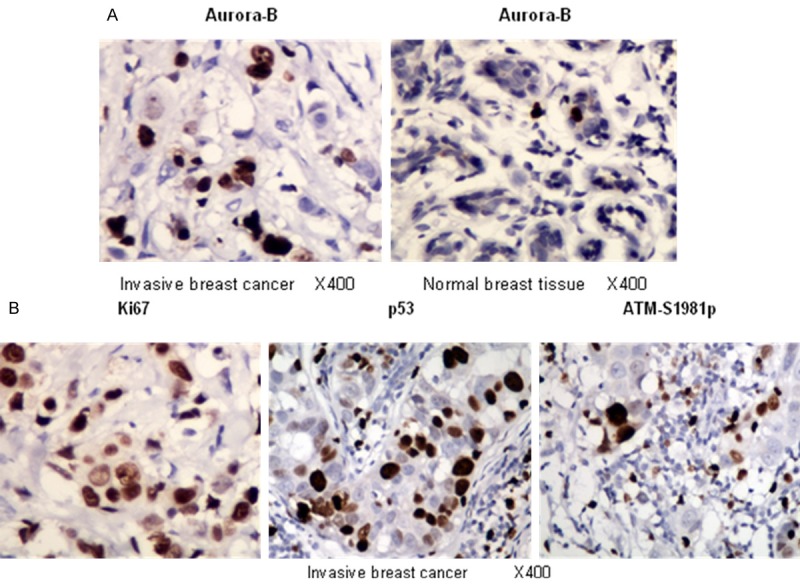
Figure 1.

Immunohistochemical study revealing representative images of invasive breast cancer and normal breast tissues with antibody of Aurora-B, nuclei with fine granular staining were counted, and (-/+) was < 5% of the cells stained, (++) was 5-10% of the cells stained, and (+++) was > 10% of the cells stained (A); and representative images of invasive breast cancer tissues stained with antibodies of Ki67, p53 and ATM-S1981p, they were scored as (-) = no positive cells, (+) = 1-10% of the cells stained, (++) = 11-50% of the cells stained, and (+++) = 51-100% of the cells stained as described previously [13] (B).
