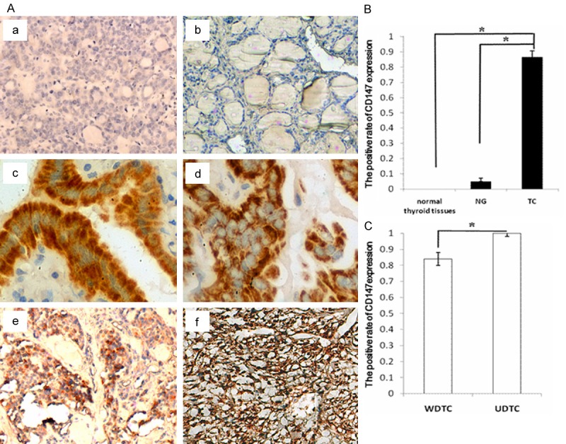
Figure 1.

Immunohistochemical staining demonstrating the correlations between CD147 expression and the pathological grading of TC tissues. A: CD147 expressed at various levels in normal thyroid tissues (a, ×200), NG (b, ×200), WDTC (c, papillary thyroid carcinoma (PTC), ×400; d, FTC, ×400) and UDTC (e, MTC, ×200; f, ATC, ×200). Staining is weak or absent in normal tissues and in NG specimens, whereas CD147 is strongly expressed in WDTC and UDTC samples. B: Comparison of the positive rate of CD147 expression. Normal thyroid tissues and NG compared with TC, *P < 0.005. C: Comparison of the positive rate of CD147 expression between WDTC and UDTC samples, *P < 0.005.
