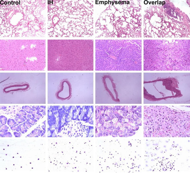
Figure 4.

Row 1~5 from the top down are histological photographs of lung, liver, right carotid artery, pancreas and cytological photographs of BALF. Histological photographs are stained with hematoxylin/eosin (H/E) and cytological photographs of BALF are stained with modified Wright-Giemsa. Photographs from lung are captured with 40 × light microscopy; liver, 100 ×; right carotid artery, 40 ×; pancreas, 400 ×; BALF, 100 ×.
