
Figure 4.
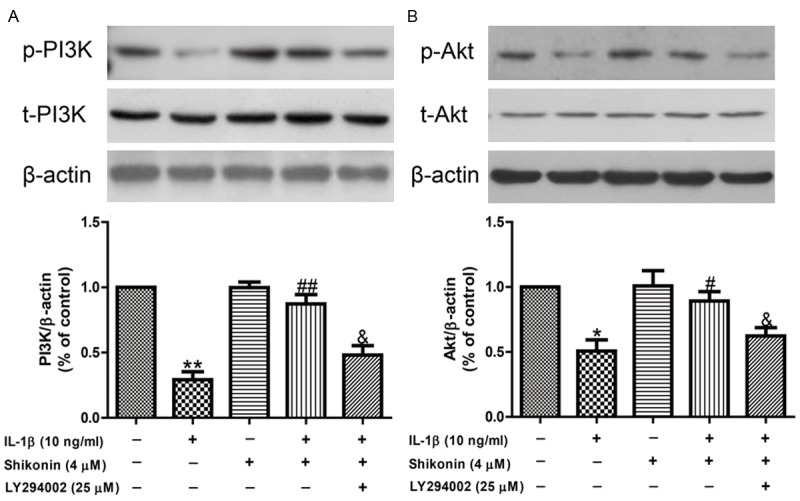
Shikonin suppressed IL-1β-induced chondrocyte apoptosis via PI3K/Akt signaling. Chondrocytes were treated with shikonin (4 μM) or shikonin (4 μM) + LY294002 (25 μM) for 2 h before 24 h IL-1β (10 ng/mL) treatment. A. Total PI3K and p-PI3K were detected by Western Blot. P-PI3K levels were normalized to t-PI3K levels. B. Total Akt and p-Akt were detected by Western Blot. P-Akt levels were normalized to t-Akt levels. All data are presented as mean ± SEM, n = 6. *P < 0.05, **P < 0.01 vs. control; #P < 0.05, ##P < 0.01 vs. IL-1β group; &P < 0.05 vs. IL-1β+ shikonin group.