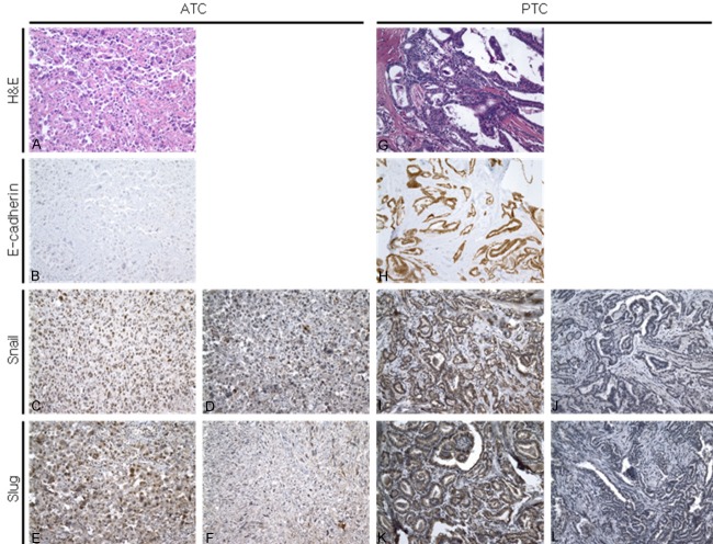
Figure 1.
The expression of EMT-related markers in ATC and PTC. ATC cells show mesenchymal features (A) and frequently show loss of E-cadherin expression (B). Snail and slug proteins are expressed in the nucleus of ATC cells and this staining is regarded as positive (C and E). Cytoplasmic staining is defined as negative (D and F). Representative hematoxylin and eosin staining (G) and staining for E-cadherin (H), snail (I, J), and slug (K, L) in PTC cases. All PTC cases show characteristic nuclear features of PTC (G) and retain E-cadherin expression (H). A small percentage of PTC cases are immunoreactive for snail (I) and slug (K), and almost all cases reveal no immunoreactivity for snail (J) and slug (L). Original magnification: ×200.

