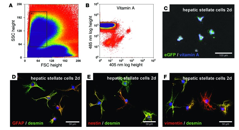
Figure 1. Characterization of freshly isolated HSCs from eGFP+ rats sorted by retinoid-dependent FACS.
HSCs were enriched by density gradient centrifugation and further purified by FACS using (A) forward (FSC) and side scatter (SSC) as well as (B) retinoid fluorescence. HSCs with (A) similar morphological characteristics (gate R1) were (B) further analyzed for their retinoid fluorescence by UV light excitation. (B) HSCs with intense retinoid fluorescence (gate) were (C) cultured for 2 days and exhibited retinoid (blue) and eGFP (green) fluorescence as assessed by blue and UV light excitation of living cells. HSCs sorted by FACS expressed (D) GFAP, (E) nestin, and (F) vimentin (red), along with (D–F) desmin (green), as determined by immunofluorescence. Cell nuclei were stained with DAPI (blue). Scale bars: 100 μm (C); 50 μm (D–F).

