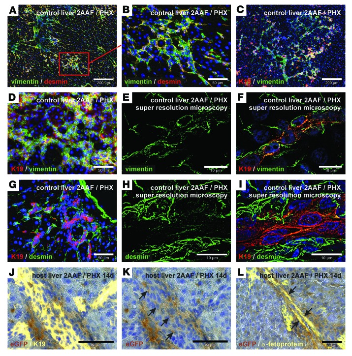
Figure 4. Transplanted eGFP+ HSCs participate in ductular reactions (2AAF/PHX model).
(A–I) Immunofluorescence of mesenchymal (vimentin and desmin) and epithelial (K19) marker proteins during ductular reactions in the liver after PHX in the presence of 2AAF. Simultaneous detection of vimentin (green) and desmin (red) in ductular structures of the portal field at (A) low and (B) high magnification. Combined immunofluorescence of K19 (red) and vimentin (green) at (C) low and (D) high magnification. Super-resolution microscopy of (E) vimentin (green) showing vimentin residues (F) in K19+ cells (red) in the center of duct-like structures. (G) Immunofluorescence of K19 (red) and desmin (green) of ductular structures within the portal field. (H and I) Desmin immunofluorescence (green) of ductular reactions was analyzed by super-resolution microscopy and (H) showed desmin residues (I) in K19+ cells (red) within the center of duct-like structures. (J–L) DAB immunohistochemistry of eGFP (brown) in WT host liver with transplanted eGFP+ HSCs. (J) Epithelial cells of ductular reactions were indicated by (J) K19 and (L) α-fetoprotein immunofluorescence (yellow). HSC-derived eGFP+ cells with (K) K19 (without immunofluorescence shown in J) or (L) α-fetoprotein are indicated by arrows. All microscopic images were taken in the portal fields of injured livers (zone 1). Scale bars: 200 μm (A and C); 50 μm (B, D, G, and J–L); 10 μm (E, F, H, and I).

