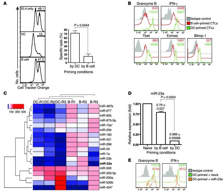
Figure 1. Identification of miR-23a as a negative correlate of CTL effector function.
(A and B) pMel-1 CTLs primed with splenic B cells or LPS-matured bone marrow–derived DCs for 4–6 days were assessed for (A) in vitro cytotoxicity at an E/T ratio of 5:1 and (B) expression of CTL effector molecules. Histograms are representative of n = 3 independent experiments, and the bar graph represents the mean ± SEM of n = 3 independent experiments. (C) After 3 days of in vitro priming by DCs or B cells, pMel-1 CTLs were isolated for miRNA expression profiling. Heat map of miRNAs differentially expressed by DC- and B cell–primed CTLs from n = 3 independent miRNA profiling experiments. Asterisks with miRNAs refer to the passenger strands of the respective miRNA species. (D) Validation of differential miR-23a expression in CTLs under the respective priming conditions with 3 additional batches of samples. Numbers and bar graph represent the mean ± SEM miR-23a expression relative to that of naive CD8+ T cells. (E) DC-primed pMel-1 CTLs were retrovirally transduced with either an empty mock vector or a miR-23a overexpression vector. Three days after transduction, CD8+GFP+ CTL effector molecule expression was assessed by flow cytometry.

