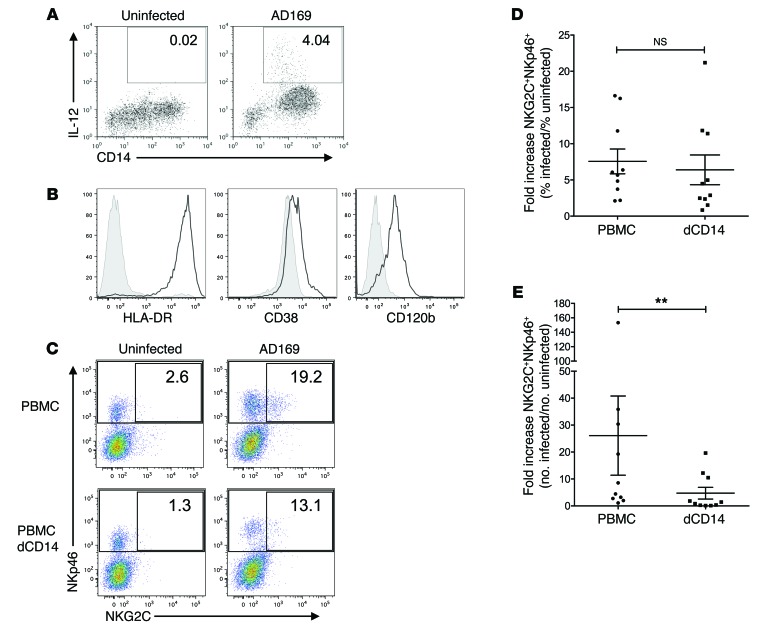
Figure 5. Impact of depletion of CD14+ cells on NKG2C+ subset expansion in HCMV-infected cocultures.
(A) PBMCs were cultured with uninfected or AD169-infected MRC-5 fibroblasts. Cells were stained for cell-surface–expressed CD14 and intracellular IL-12 at 36 hours p.i. Displayed are cells within the monocyte gate. Numbers indicate the percentages of IL-12–producing monocytes (1 representative donor out of 6 is shown). (B) Monocytes were stained for HLA-DR, CD38, CD120b (black line), or the respective isotype controls (shaded). Histograms are gated on IL-12+CD14+ cells (1 representative donor out of 3 is shown). (C) PBMCs or PBMCs depleted of CD14+ cells (dCD14) were cocultured with uninfected or infected fibroblasts. At the end of the coculture, cells were stained for NKp46, NKG2C, and CD3 and analyzed by flow cytometry. Dot plots were gated on live CD3– cells. Numbers indicate the percentages of NKG2C+ cells among all NKp46+CD3–cells (1 representative donor out of 10 is shown). (D and E) Summary of cocultures of PBMC or PBMC with CD14 depletion (dCD14) is shown. Depicted are the fold increases of percentage (D) and absolute numbers (E) of NKG2C+ among NKp46+ cells in infected versus uninfected cocultures. Wilcoxon matched pairs signed-rank test: **P = 0.002; n = 10; error bars indicate ± SEM.

