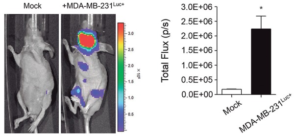
Figure 1. Mouse model for metastatic breast cancer to the mandible. The growth and part of the metastatic spread (left) was detected by bioluminescence imaging after the injection of cancer cells. The formed metastases were quantified by measuring total photon flux per second (right). Data are expressed as the mean±standard error (SEM). *P<0.001 versus Mock group.

