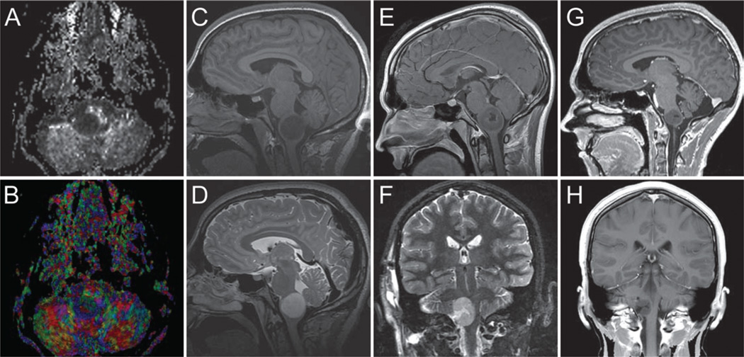
Figure 2.
Images obtained in a 17-year-old boy who presented with medullary WHO Grade II astrocytoma. Three separate time points are selected for demonstration. A and B: Preoperative fractional anisotropy grayscale and color maps showing baseline imaging. C and D: Preoperative sagittal T1-weighted noncontrast (C) and T2-weighted MR images (D). E and F: Postoperative, preirradiation sagittal T1-weighted MR image obtained after contrast administration (E) and coronal T2-weighted MR image (F) obtained 1 week later. G and H: Postirradiation sagittal (G) and coronal (H) T1-weighted MR images obtained after contrast administration 21 months later.

