Figure 1.
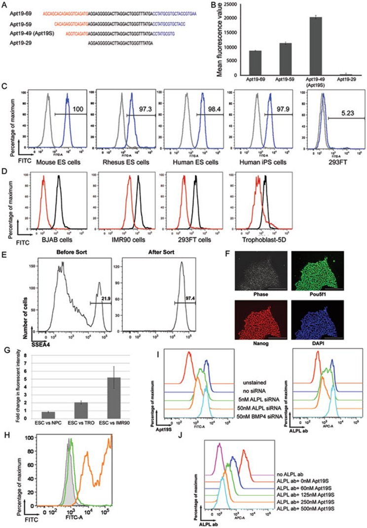
Characterization and target identification of a DNA aptamer for PSCs. (A) Diagram showing the location of truncations made in the constant regions of Apt19. (B) The affinity of aptamers in A towards hESC was measured with flow cytometry (error bar, SEM; n = 3). (C) Different types of PSCs were incubated with FITC-labeled Apt19S (blue) or Apt19S-SC (grey). The fluorescent intensities of the cells were then measured by flow cytometry. Human 293FT cells were used as a negative control. (D) Different types of differentiated human cell types were incubated with FITC-labeled Apt19S (red) and analyzed by flow cytometry. Human ESCs incubated with Apt19S were used as a positive control (black). (E) A mixture of hESCs and human foreskin fibroblast cells were incubated with biotin-labeled Apt19S and FITC-labeled SSEA4 antibody. After incubation, Apt19S was separated from unbound cells by streptavidin-labeled magnetic beads. The fluorescence profiles of the cell mixture before streptavidin beads separation (left) and cells bound to the beads (right) were analyzed by flow cytometry. The number in each plot shows the percentage of SSEA4+ hESCs in the cell mixture. (F) Beads-bound cells in E were expanded in culture and stained for pluripotency markers Pou5f1 and Nanog. Cell nuclei were labeled with DAPI (scale bar, 200 μm). (G) Fluorescently labeled Apt19S was incubated with H1 ESCs, NPCs, TRO cells and IMR90 fibroblast cells. The fluorescent intensity of each cell type was measured by flow cytometry after incubation and presented as the fold changes between H1 cells and other cell types (error bar, SEM; n = 3). (H) Fluorescently labeled Apt19S was incubated with 293FT cells (grey shade), 293FT cells overexpressing ALPL (orange line) or PROM1 (green line). The fluorescent profiles were then measured by flow cytometry. (I) The binding of fluorescently labeled Apt19S (left panel) or an ALPL antibody (right panel) to hESCs treated with siRNAs against either ALPL or BMP4 was analyzed by flow cytometry. (J) Binding of a fluorescently labeled ALPL antibody to hESCs in the presence of increasing amounts of Apt19S was analyzed by flow cytometry.

