Figure 4.

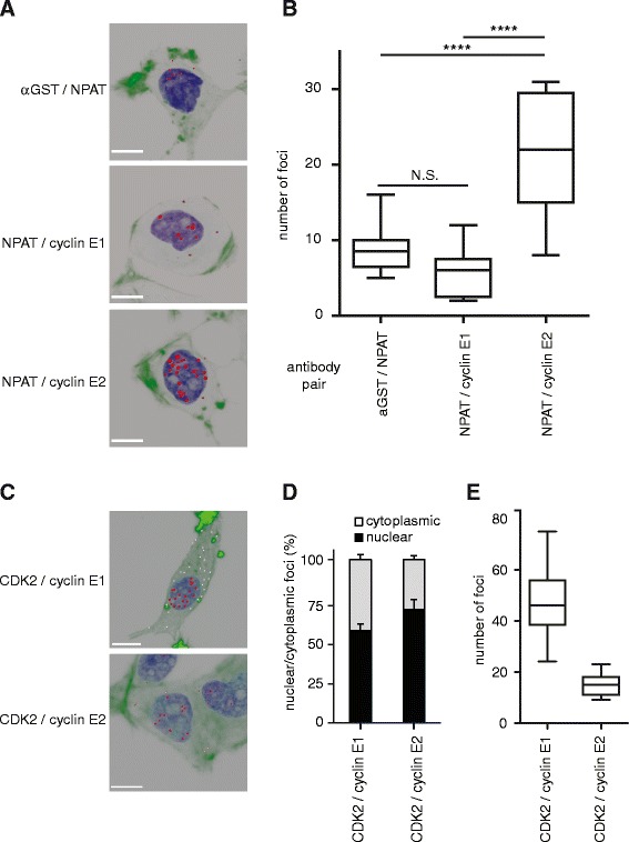
Cyclin E2, but not cyclin E1, co-localises with NPAT in T-47D cells by PLA. A. Proximity Ligation Assay (PLA) for cyclin E1/NPAT (antibodies: cyclin E1 – Epitomics; NPAT – 27) and cyclin E2/NPAT (antibodies: cyclin E2 – Epitomics; NPAT – 27). Images are 3-D rendered serially stacked confocal images assembled with Imaris software. NPAT/αGST staining was performed as a negative control (antibodies: NPAT – 27, αGST – [23]). Representative cells are shown, scale bars = 10μm. B. Quantitation of A. where number of foci were quantitated from 10-15 cells per antibody pair. Statistical significance was calculated with one-way ANOVA and Tukey’s multiple comparisons, where N.S. indicates not significant and **** indicates P < 0.0001. Data pooled from duplicate experiments. C. Cyclin E1/CDK2 (cyclin E1- HE12, CDK2 – M2) and cyclin E2/CDK2 (cyclin E2 – Epitomics, CDK2 – D12) PLA were performed as positive controls. Representative cells are shown with nuclear foci pseudocoloured in red, and cytoplasmic foci pseudocoloured in white. Scale bars = 10μm. D./E. Quantitation of C. including relative nuclear/cytoplasmic foci (D.) and total foci (E.). Data pooled from duplicate experiments.
