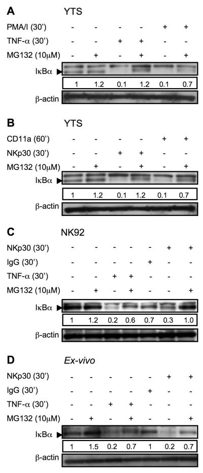
Figure 3. Inhibition of activation induced IκBα degradation in YTS, NK92 and ex-vivo NK cells by MG132.
YTS cells were pretreated with MG132 for 30 min, restimulated with TNF-α or PMA/I (A), anti-CD11a or anti-NKp30 (B). NK 92 (C) and ex vivo NK cells (D) were pretreated with MG132 for 30 min, restimulated with TNF or anti-NKp30 for 30 min and then lysed. Lysates were separated, transferred and membranes probed with anti-IκBα rabbit pAb (~33KD), as demonstrated by the arrowhead. Membranes were then stripped and reprobed with anti-βactin rabbit pAb (~38KD) as a control for protein loading. A nonspecific band above the IκBα band was found with some lots of the pAb. Numbers below each IκBα blot represent the ratio of the band intensity to the 30 min band intensity for IκBα. Representative results of three independent experiments are shown.

