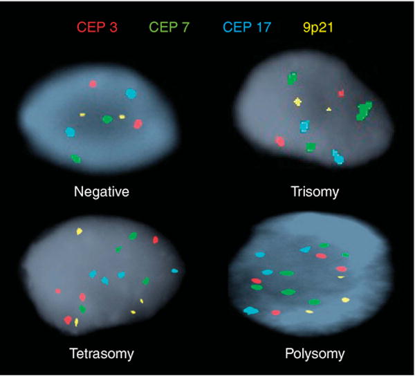
Figure 1.

Subtypes of fluorescence in situ hybridization. Individual cells showing different fluorescence in situ hybridization results using CEP to chromosomes 3 (red), 7 (green), 17 (aqua) and a locus specific probe targeting 9p21 (gold). Negative (two copies of each probe); Trisomy 7 (at least 10 cells with ≥3 CEP 7 signals and ≤2 signals of the other probes); Tetrasomy (at least 10 cells with four signals for all four probes); Polysomy (at least five cells showing ≥3 signals for at least two of the four probes). CEP, centromere enumeration probe.
