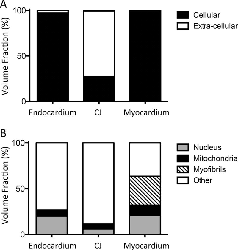
Figure 5. Cellular and sub-cellular volume fractions in the endocardium, cardiac jelly, and myocardium.

Reconstructed and aligned 3D image stacks of the cardiac layers were manually segmented. The volume of each segmented region was ascertained, allowing for a quantitative comparison of volume fractions. (A) Cellular and extra-cellular volume fractions for each layer of the cardiac wall. As expected, only the cardiac jelly layer contained a significant portion of extra-cellular tissue. (B) Volume fractions of sub-cellular structures (nuclei, mitochondria, myofibrils) within the cells.
