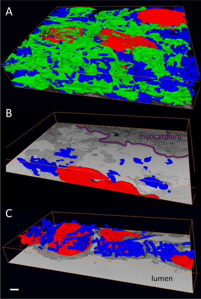
Figure 6. Isosurface renderings of 3D segmentations within each of the three layers of the OFT wall.

(A) Myocardium; (B) cardiac jelly; (C) endocardium. The segmentations show sub-cellular structures within the cells in each layer: nuclei (red), mitchondria (blue) and myofibrils (green). Note that the myofibrils, which are active contractile proteins, are only present in the myocardium. Also, a comparison of the panels demonstrates the densely packed nature of the myocardium and the mostly extracellular nature of the cardiac jelly. The endocardium consists of tightly packed, rounded cells that line the OFT lumen. Scale, 1 μm.
