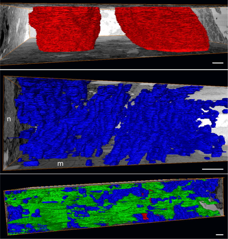
Figure 7. Segmented sub-cellular structures of the early embryonic myocardium.

(A) Nuclei (red) are shown spanning the depth of this FIB-SEM dataset. (B) Mitochondrial (blue) organization is apparent even at this early stage (HH24), and is evidenced by the stacked appearance and consistent directionality. A nucleus (n) and myofibrils (m) are evident on the bounding orthoslices. (C) Myofibrils (green) lack organization at this stage, appearing in random orientations and directions throughout the tissue. Scale, 1 μm.
