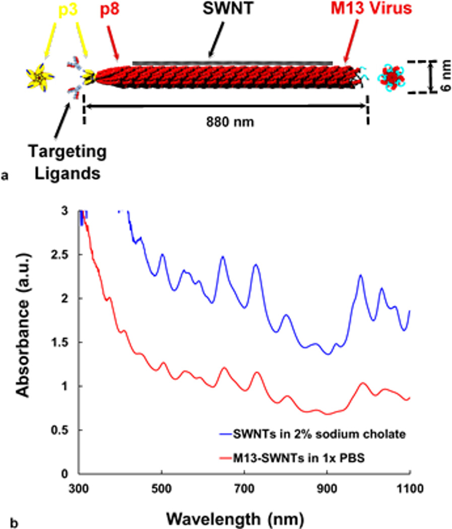
Figure 1. Schematic of the SWNT probe and its absorbance spectrum.
(a) Structure of the SWNT probe. M13 is a filamentous, cylindrical bacteriophage, rendered in red, with typical dimensions ~880nm in length and ~6nm in diameter. Of interest is the major coat protein, p8, which can be used to longitudinally functionalize SWNTs, shown in black; and the minor coat protein, p3, which can be used to bind targeting ligands such as antibodies. (b) Absorbance spectra of aqueous-dispersed SWNTs; using 2 wt.% sodium cholate, an organic surfactant (blue curve), and after surfactant-exchange to form the M13-SWNT complex (red curve). The characteristic peaks are maintained, although there is slight red shift in the M13-SWNT spectrum.

