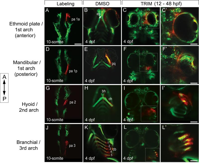
Figure 2. Ectopic cartilages were derived from malformed ceratobranchial as a result of the failure in midline convergence of CNC cells.
Photoconversion labeling of CNC cells in sox10:kaede followed from 10-somite stage (A,D,G,J) to 4 dpf, after treatment with DMSO (B,E,H,K, ventral view) and TRIM (C,F,I,L, ventral view). Anterior is to the top.
(A-C) CNC cells anterior of the eye were fated to the first PA (A, pa1a) and contributed to ep (B, arrow). In TRIM-exposed embryos, the anterior CNC cells failed to converge and condense to the midline (C). C’, enlarged view of the dotted area in C in dorsal focus.
(D-F) The posterior population of the first PA (D, pa 1p) normally populates the lower jaw structures (m, pq) (E). After TRIM treatment, the cells were sequestered in a lateralized domain posterior to the eyes (F). F’, enlarged view of the dotted area in F in dorsal.
(G-I) CNC cells in PA 2 (G) normally populate the bh, ch (H). After TRIM treatment, the cells in PA2 failed to converge in the midline and were stuck in the lateral position posterior to the eyes (I). I’, enlarged view of the dotted area in I in lateral.
(J-L) CNC cells that give rise to the third and posterior PA (J, pa3) formed paired segmented ceratobranchials at 4 dpf (K, cb 1-5). When these cells were followed in TRIM-treated embryos, they remained lateralized without midline convergence, thus failed to form the ceratobranchials (L). L’, enlarged view of the dotted area in L in lateral.
bh, basihyal; ch, ceratohyal; cb, ceratobranchial; ep, ethmoid plate; pq, palatoquadrate. Scalebars: A-L, 50 μm; C’, F’, I’, L’, 20 μm.

