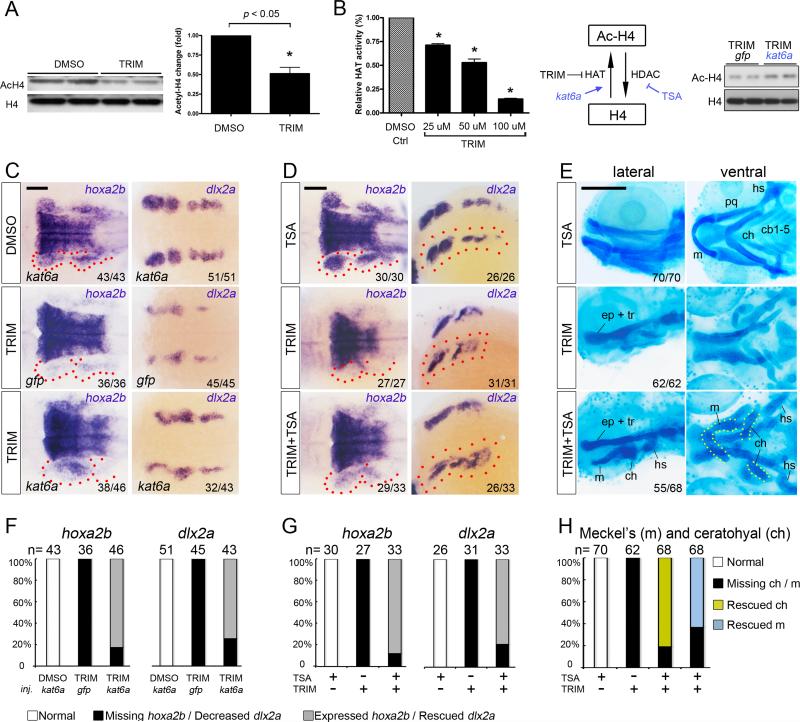
Figure 5. Potentiating histone acetylation counteracts TRIM-induced craniofacial defect.
(A) Impaired histone H4 acetylation was detected in TRIM-treated embryos compared to DMSO control. Quantification data is presented as mean ± s.e.m. *P < 0.05.
(B) Treatment with TRIM inhibited the activity of histone acetyltransferase in a dose-dependent manner. Injection of kat6a mRNA partially counteracts TRIM-induced hypoacetylation.
(C) Overexpression of kat6a mRNA antagonizes TRIM-induced genetic defect by partially rescuing expression of CNC marker hoxa2b and dlx2a in PA (24 hpf; middle versus bottom panels). Scalebar 100 μm.
(D) Chemical potentiation of histone acetylation by TSA partially restored TRIM-induced hoxa2b and dlx2a depletion in PA (24 hpf; middle versus bottom panels). Scalebar 100 μm.
(E) TRIM-induced skeletal abrogation was partially rescued by TSA co-treatment, as demonstrated by formation of the 1st and 2nd PA cartilage structures, including the Meckel's cartilage (m), ceratohyal (ch), and hyosymplectic (hs) (96 hpf, middle versus bottom panels). Scalebar 250 μm.
(F, G, H) Quantification of the rescuing phenotype observed in C, D, E, respectively. The statistical data are presented as percentage of embryos with corresponding phenotype.

