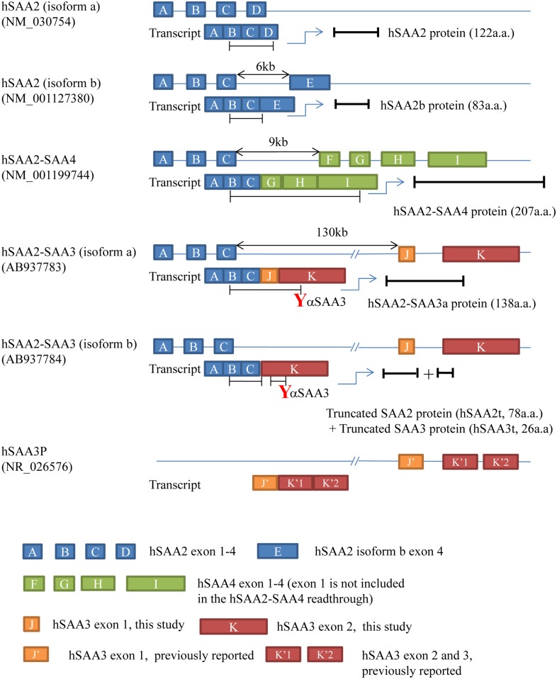
Fig 1. Structures of hSAA2-SAA3 readthroughs.
A-K stand for exons in the hSAA2, hSAA3, or hSAA4 gene. Human SAA3 exon 1 (J) identified in the present study was 52 bp shorter than that previously reported (J’). Human SAA3 exon 2 (K) in the present study contained intron between hSAA3 exon 2 (K’1) and exon 3 (K’2). The 5’ edge of (K) was identical to that of (K’1), as was the 3’ edge of (K) to that of (K’2). The bars under the transcripts represent open reading frames. As displayed in the figure, our hSAA3 antibodies recognize the C-terminal region of hSAA3.

