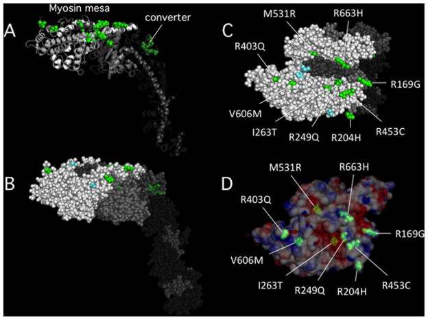
Figure 6. The locations of 15 HCM mutations in the myosin catalytic domain.
(A) Cartoon image view showing two groups of HCM mutations. Four mutations are in the converter domain (dark green) and 11 are on or very near the myosin mesa (bright green). Fourteen of the mutations were chosen because they have been well-documented to be the cause of HCM in families carrying these mutations. The fifteenth is M531R, which, although only documented in one family, is a left ventricular non-compaction mutant myosin that appears to be hypercontractile in our studies. (B) The same view as in (A) except in all sphere representation. (C) The top view of the mesa showing that nine of the 11 mutations are on the mesa surface (the other two are just below the surface). (D) The same view as in (C) except the surface charge distribution is shown. M531R and I263T are slightly below the surface in acidic pockets, while the remainder of the mutations are right at the surface and most are arginine residues, 5 of which form a particularly large domain of positive charge on the lower right (see also Figure 3C, small dashed oval).

