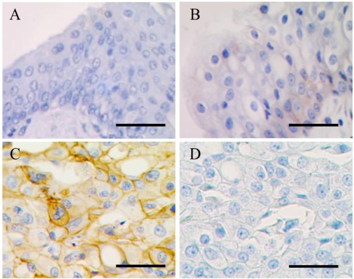
Fig. 1.
Images of immunohistochemistry. Normal (A), polypoid cystitis (B) and TCC (C) epithelial cells show no, weak and intense immunohistochemical staining at the cell membrane, respectively. In a negative control study (D), no significant staining was observed at the neoplastic cell membrane in TCC. Bar=100 µm

