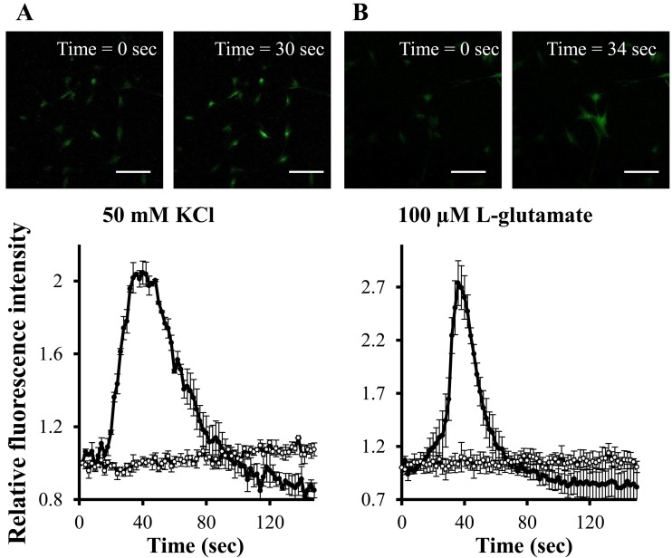
Fig. 5.
K+- and L-glutamate-induced mobilization of Ca2+ in neuron-like cells derived from canine BMSCs after incubation with (closed circles) and without (open circles) bFGF (100 ng/ml). The cells were stimulated with either 50 mM KCl (A) or 100 µM L-glutamate (B). Images of Ca2+ response to KCl or L-glutamate in the fluorescent dye-loaded cells treated with bFGF were displayed in the upper panel. Green fluorescence shows the changes in intracellular Ca2+ concentration, indicating neuronal activation. Changes in intracellular Ca2+ concentration are displayed in the bottom panel. The scale bar is 200 µm.

