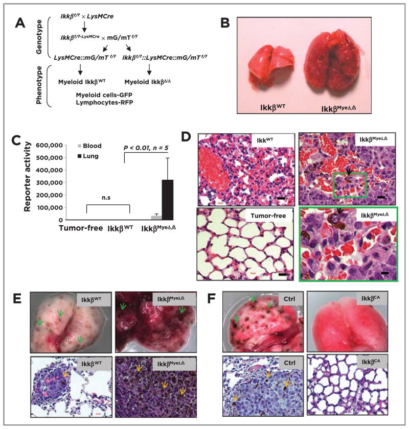
Figure 1.
Deletion of Ikkβ in myeloid cells leads to protumorigenic immunity. A, generation of mice with myeloid Ikkβ deletion. Ikkβf/f mice (C57BL6) crossed with myeloid specific LysMCre mice (C57BL6) and mG/mT mice (C57BL6). Cre+ myeloid cells express GFP, whereas other Cre+ cells (e.g., lymphocytes) express Tomato red protein. B, BrafV600E/Pten−/− melanoma cells form melanoma lung lesions in IkkβMyeΔ/Δ mice, but not in IkkβWT mice. C, quantitation of lung melanoma lesions based on Gluc activity (mean ± SEM) in the blood or lung 4 weeks after G-luc-BrafV600E/Pten−/− cells were i.v. injected into IkkβΔ/Δ or IkkβWT C57B/6 mice or tumor-free IkkβWT mice. D, histology of melanoma lesions in lungs from C based on H&E staining. Arrows, melanoma cells in lungs of IkkβmyeΔ/Δ mice. E, myeloid IKKβ deletion enhances B16F0-Gluc melanoma growth in lung in syngeneic IkkβMyeΔ/Δ mice versus littermate IkkβWT mice. Twenty days after i.v. injection of tumor cells, lungs were perfused and photographed (top), or fixed and stained with H&E (bottom). F, B16F0 melanoma growth in C57BL6 myeloid IKKβCA mice and littermate control mice (single transgene cfms-rtTA or TetOn cIKKβ) received B16F0-Gluc melanoma cells i.v. and transgene was induced with doxycycline. After 20 days, lungs were collected for photography (top) and H&E staining (bottom). Arrows, the pigmented melanoma lesion. Scale bar, 50 μm.

