Figure 4.
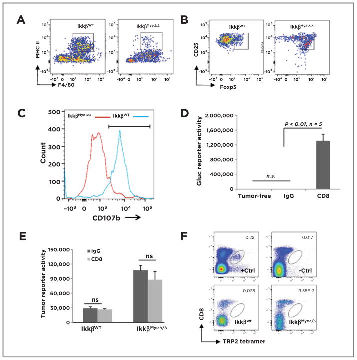
Myeloid IKKβ mediates macrophage and CD8+cytotoxicity. A, C57Bl/6 IkkβWT or IkkβMyeΔ/Δ mice were i.v. injected with BrafV600E/Pten−/− cells. After 24 hours, GFP+ lung macrophages were stained for F4/80 and MHCII and analyzed by FACS. B, BrafV600E/Pten−/− cells were injected i.v. into mice carrying IkkβWt or IkkβMyeΔ/Δ myeloid cells. Pulmonary Tomato-RFP CD4+ T cells double-positive for CD25 and Foxp3 were analyzed by FACS 3 days after injection. C, using the protocol described in B, lung Tomato-RFP lymphocytes positive for both CD8 and CD107b were evaluated by FACS. D, CD8+ cells were depleted and after 3 weeks, lung tumor burden was analyzed by Gluc activity. E, CD8+ T cells were depleted 3 days before subcutaneous implantation of 5 × 10⁴ Gluc-B16F0 melanoma cells. CD8 or control antibody injections continued 16 days before tumor burden was assessed by Gluc activity assay. F, tetramer analysis of CD8+T cells infiltrating syngeneic melanoma tumor. IkkβWt or IkkβMyeΔ/Δ mice received B16F0 melanoma cells (5 × 10⁴) i.v. After 16 days, cells from the lungs of these mice were stained with PerCP-Cy5.5–conjugated CD8 antibody and APC-labeled tetramer with monocyte-derived TRP2 (SVYDFFVWL) peptide and analyzed FACS. +Ctrl, positive control cells from splenocytes of TRP2-immunized mouse; −Ctrl, negative control cells from splenocytes of nonimmunized mouse.

