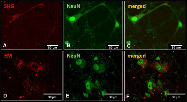
Figure 10.

Immunohistochemical detection of neuronal phenotype of transduced cells. Co-localization of SHG (A) or EM2 (D) transduced cells expressing reporter gene mRFP (red) with neuronal marker (NeuN, green)) (B,E). Overlapping images show the presence of punctate staining in the cytoplasm of neuronal cells (C,F).
