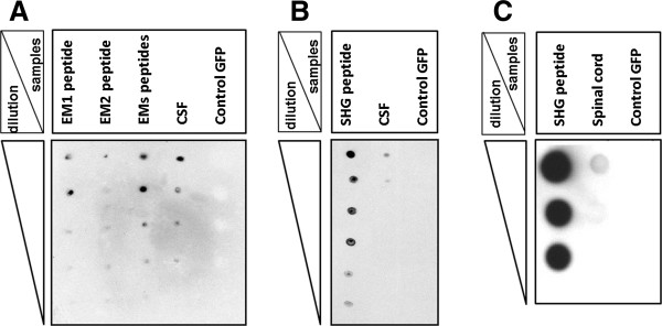
Figure 12.

Immunodot-blot analysis of CSF and spinal cord of treated rats. Immunodot-blot assay of CSF from the GFP control SCI animals and SCI animals injected by a mixture of viral vectors encoding SHG and EMs. No reactivity was seen in the CSF of GFP control SCI animals. The amount of each peptide spotted is indicated in the column on the left side of each panel. A: reactivity of CSF with endomorphin 1 and 2 antibody, B: reactivity of CSF with SHG antibody. C: detection of SHG in the spinal cord homogenate from EMs + SHG treated animals.
