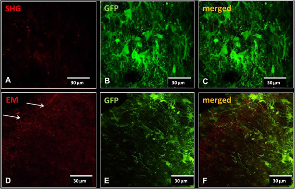
Figure 9.

Injection of control vector expressing GFP only. No SHG signal was detected in spinal cords of animals receiving control GFP vector injection (A). Staining with EM1/2 in these animals revealed punctate staining of the endogenous EM in the dorsal horn (arrows in D). B and E show the GFP fluorescence in cell bodies and processes in the same sections (A and D, respectively). C and F are merged images to reveal any co-localization.
