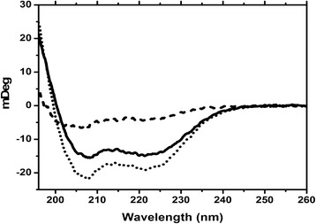
Figure 7.

Circular dichroism spectra of uncomplexed and complexed CaM. The CD spectra of CaM were recorded in the presence of Ca2+ (solid line) and Ca2+ and Orai-CMBD (dotted line). Their differential spectrum (dashed line) presents the structure of Orai-CMBD in the complex assuming no CaM structural change upon ligand binding. Note that the CD unit was intentionally shown in degrees of light rotation because the CD spectra of CaM and the complexed Orai-CBMD have a very similar mean residual ellipticity.
