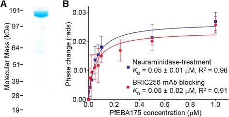
Figure 3.

Backscattering interferometry detects the PfEBA175-GYPA interaction on intact unmodified erythrocytes. (A) The entire ectodomain of PfEBA175 was expressed and purified as a Cd4-tagged monomer and resolved under reducing conditions by SDS-PAGE. Coomassie staining revealed a single band of the expected size (185 kDa). (B) Quantitation of PfEBA175 binding to intact unmodified erythrocytes at equilibrium using BSI. Erythrocytes that were either neuraminidase-treated (blue squares) or pre-coated with anti-GYPA monoclonal antibody BRIC256 (red circles) were used as control samples. Data points are means ± SD from three independent experiments.
