Figure 1.

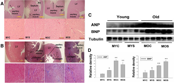
Representative histological cross sections of the left ventricle (LV) stained with hematoxylin-eosin stain and Masson’s trichrome staining. Substantial left ventricles remodeling in old age heart and secondhand smoke exposure. (A) Morphological features of the left ventricle of hematoxylin-eosin stained. Morphometric samples are at x100 magnification (up-panel). Morphometric samples are at x400 magnification) (down-panel). The arrow points to the papillary muscle. (B) Histological examination of LV fibrosis by Masson’s trichrome staining. Morphological features of left ventricular hematoxylin-eosin stained. Morphometric samples are at x100 magnification (up-panel). Morphometric samples are at x400 magnification) (down-panel). The long arrow points to the papillary muscle, short arrow points to collagen. (C) western blotting for ANP and BNP protein in MYC, MYS, MOC and MOS groups from the indicated left ventricular extracts. (D). Statistical analysis ANP and BNP protein expression levels in MYC, MYS, MOC and MOS groups. Data are means ± SD. *P < 0.05, **P < 0.01 significantly statistical differences vs. MYC group (two-way ANOVA). MYC; male young control, MYS; male young SHS exposure, MOC; male old control, MOS; male old SHS exposure.
