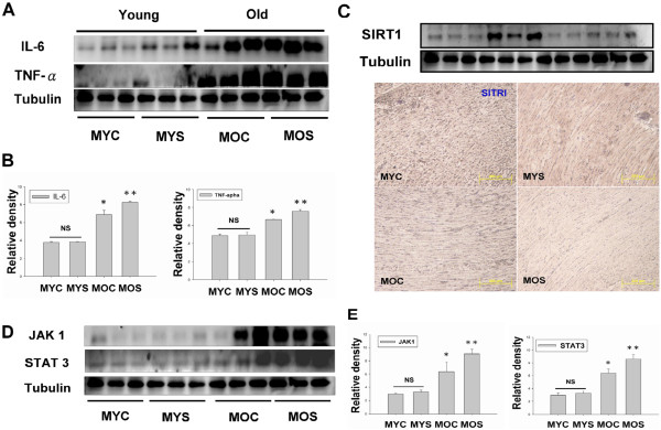
Figure 3.

Inflammatory cytokines, IL-6, TNFα, JAK1 and STAT3 mediated eccentric left ventricular hypertrophy in aging and aged SHS exposure hamsters. (A). Western blot analysis of Inflammatory protein, IL-6 and TNFα, expression in MYC, MYS, MOC and MOS groups. (B). Quantification of densitometry analysis of IL-6 and TNFα protein expression levels. Statistical analysis of IL-6 and TNFα protein expression levels in MYC, MYS, MOC and MOS groups. All data are presented as means ± SD. *p < 0.05, **p < 0.01 significant statistical differences compared with male young control (MYC). (C). Western blot and immunohistochemical analysis detection of SIRT1 in the four experimental groups were measured. (D). Western blot analysis of cytokines, JAK1 and STAT3 protein expression in MYC, MYS, MOC and MOS groups. (E). Quantification of densitometry analysis of JAK1 and STAT3 protein expression levels. All data are presented as means ± SD. *p < 0.05, **p < 0.01 significant statistical differences compared with male young control (MYC).
