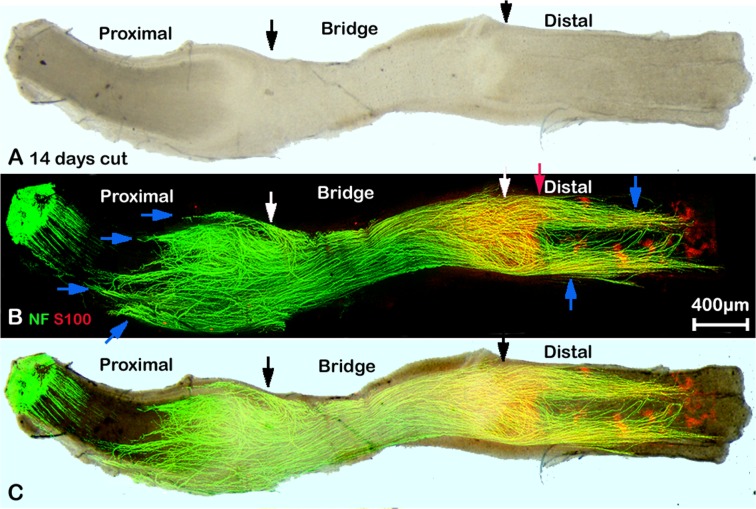
Fig 5. Whole mount staining of transected sciatic nerve preparation after 14 days injury showing misdirected regrowing axons.
A: phase image showing the structure of transected sciatic nerve, black arrows mark the proximal (left) and distal (right) nerve stumps. B: neurofilament (NF) and S100β (S100) antibodies whole mount staining show regenerating axons in the nerve bridge and misdirected regrowing axons in both proximal and distal nerve stumps, blue arrows indicate large axon bundles that have turned 180 degrees and have grown back along the outside of the proximal nerve stump. Axons that are growing outside of the distal part of the nerve (blue arrows) can be seen more clearly at 14 days after injury. White arrows mark the proximal (left) and distal (right) nerve stumps. Red arrow shows the distance of neurofilament/S100β antibody penetration within the distal part of the nerve. C: merged image of panels A and B.

