
Figure 3.
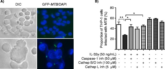
IL-32 reduces the proportion of MTB -infected THP-1 cells and this effect is partly dependent on caspase-1, cathepsins B/D, and cathepsin L activity. Differentiated THP-1 cells were infected with GFP-MTB H37Rv with the indicated conditions for four days and the proportions of MTB-infected cells were quantified by fluorescent microscopy. A) Representative DIC and fluorescent microphotograph of control THP-1 cells infected with GFP-labeled MTB H37Rv at lower magnification (400×, top) and higher magnification (600×, below). B) The proportion of THP-1 cells infected with GFP-MTB H37Rv was quantified after four days of culture for the indicated conditions. Data shown are the mean ± SEM of two independent experiments, each performed in duplicate. *p < 0.05, **p < 0.01.