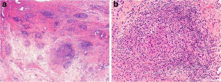
Fig. 5.
Biopsy specimen of the right wrist. Low-power view reveals a vaguely nodular process in a background of synovial and fibrovascular tissue (a). At high magnification, the nodules are comprised of clusters of multinucleated giant cells surrounded by a rim of lymphocytes, plasma cells, and histiocytes (b). These morphologic features are consistent with non-necrotizing granulomatous inflammation. Stains for microorganisms (Grocott’s methenamine silver, acid fast bacilli) were negative (not shown), and no foreign body material was appreciated under polarized light. Magnification ×40 (a), ×200 (b). Courtesy of Dr. Anja C. Roden, Mayo Clinic Rochester, MN

