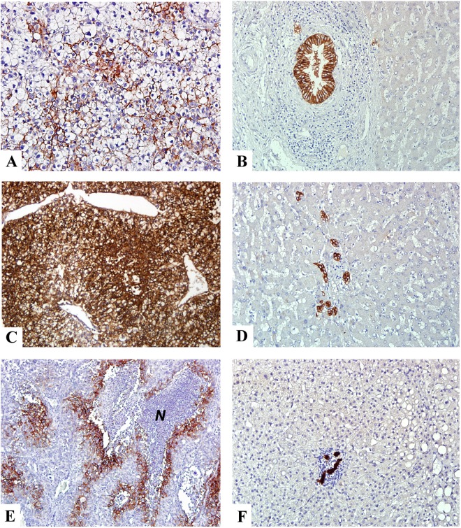
Fig 1. Expression of CA-IX protein in non-cancerous liver parenchyma and HCC.
(A and C) Immunohistochemical staining showed heterogeneous and diffuse membranous/cytoplasmic expression of CA-IX in HCC. (E) In the region of tumor necrosis, the viable tumor cells surrounding the necrotic area (N) exhibited strong CA-IX expression. (B, D, F) B, D, and F were the nontumorous counterpart of A, C, and E, respectively. They exhibited strong staining in the bile duct epithelial cells in the portal area but not in the hepatocytes or the mesenchymal cells. A-F x200 (original magnification).

