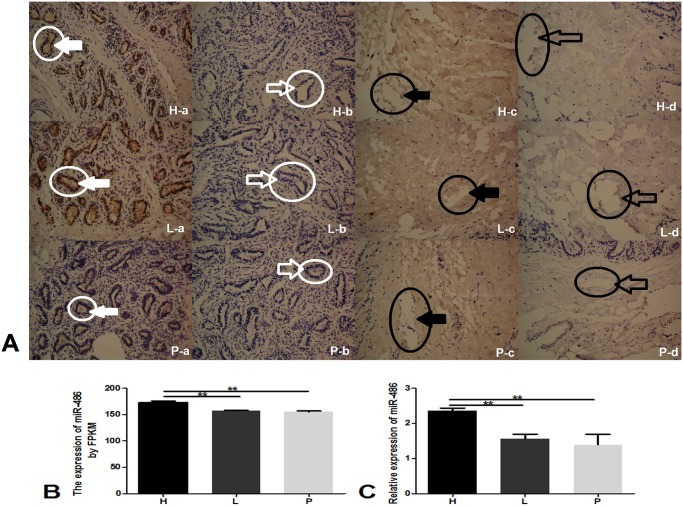
Fig 1. The expression of miR-486 in different milk qualities bovine mammary glands.
A: ISH showing the localization of miR-486 in the mammary gland. H: lactation stage with high milk quality; L: lactation stage with low milk quality; P: pregnancy stage. (a) Mammary gland tissue incubated with a DIG-labeled locked nucleic acid probe against miR-486 (probe positive). And the white circle with solid white arrow indicates miR-486 positive probe in mammary gland tissue. (b) Mammary gland tissue incubated with a negative control probe (probe negative). And the white circle with feint arrow indicates miR-486 negative probe in mammary gland tissue. (c) Adipose tissue incubated with a DIG-labeled locked nucleic acid probe against miR-486 (fat positive). And the black circle with solid black arrow indicates miR-486 positive probe in adipose tissue. (d) Adipose tissue incubated with a negative control probe. And the black circle with feint black arrow indicates miR-486 negative probe in adipose tissue. B: The results of small RNA sequencing for miR-486 (fat negative). C: Analysis of the relative expression of miR-486 in bovine glandular tissue by qRT-PCR. n = three cows in every group. qRT-PCR reactions were performed in triplicate to detect miR-486. **P<0.01.

