Fig. 3.
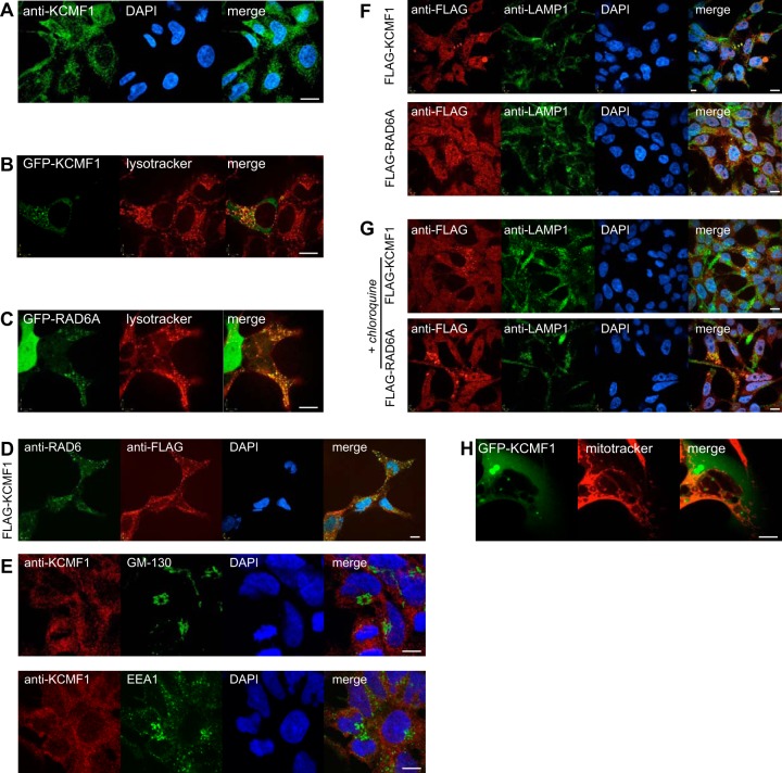
KCMF1 links RAD6 to vesicle dynamics. A, Immunofluorescence was conducted in untransfected HEK 293 cells using an antibody directed against KCMF1. DNA stained with DAPI (blue). All scale bars 10 μm. B, Colocalization of GFP-KCMF1 with Lysotracker red. C, Colocalization of GFP-RAD6A with Lysotracker red. D, Colocalization of endogenous RAD6 (green) with Flag-KCMF1 (red) in 293 T-REx cells. E, Endogenous KCMF1 does not colocalize with GM-130 (top) or EEA1 (bottom). F, Colocalization of Flag-KCMF1 (top panel, red) and Flag-RAD6A (bottom panel, red) with the late endosome/lysosome marker LAMP1 (green). G, cells treated with the lysosome inhibitor chloroquine were analyzed as in F. H, GFP-KCMF1 does not colocalize with the mitochondrial marker Mitotracker (red).

