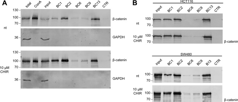
Fig. 4.
Nanobodies bind endogenous β-catenin. A, HEK293T cells were either left untreated (nt) or were incubated with CHIR (GSK3β-inhibitor) for 24 h. Cells were lysed under native conditions and membrane-bound fraction was depleted using Concanavalin A (ConA)-beads. Depleted supernatants were adjusted to 2 mg/ml and incubated with equal amounts of immobilized Nbs. Bound protein fractions were subjected to SDS-PAGE followed by immunoblot analysis using antibodies specific for β-catenin and GAPDH. Total: 0.5% of total lysate; ConA: 2.5% of ConA-bound fraction; input: 0.5% of ConA-depleted supernatant; BC1–BC13: 10% of Nb-bound fraction, CTR: 10% of bound fraction of a nonrelated Nb. Shown are representative blots of three independent experiments. B, Precipitation of β-catenin with immobilized Nbs as described in A from membrane-depleted lysates derived from HCT116 cells (upper panel) or SW480 cells (lower panel). Cells were either left untreated (nt) or were incubated with CHIR for 24 h. Immunoblot analysis of input and bound fraction with anti-β-catenin antibody is shown.

