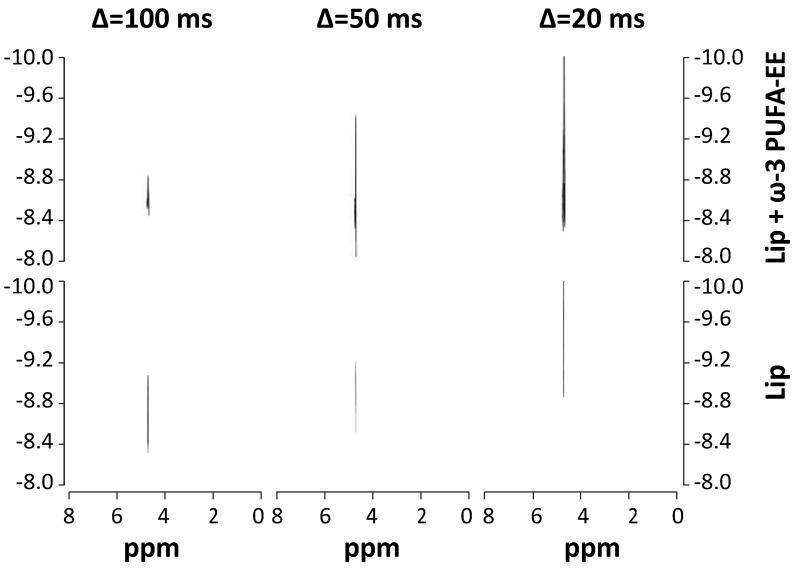
Figure 3.
Characterization of PUFA-EE nanogoticule localization. 1H NMR DOSY (400,7 MHz, 22 °C) spectra of translational water diffusion in liposomal suspensions at different diffusion times Δ. Spectra were obtained from liposomal suspensions loaded (top panels) or not (bottom panels) with ω-3 PUFA-EE, with Δ of 100 ms (left), 50 ms (center) and 20 ms (right). Note that the presence of ω-3 PUFA-EE does not decrease significantly the water ADC at any of the diffusion times investigated, suggesting primarily an intraluminal location of the goticules.

