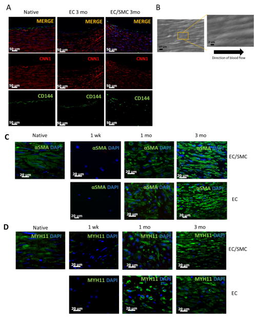
Figure 3. Immunohistochemistry and SEM.
(A) Immunostaining of tissue sections of explanted grafts (3 mo) or native arteries for CD144 (green) or CNN1 (green). Nuclei were counterstained with DAPI (blue). Bar= 50 μm. (B) SEM en-face of explanted tissue showing confluent endothelial monolayer aligned in the direction of blood flow. (C) Immuno-staining for αSMA (green) or (D) MYH11 (green) of graft sections at the indicated times. Native artery served as control. Nuclei were counterstained for DAPI (blue). Bar= 20 μm.

