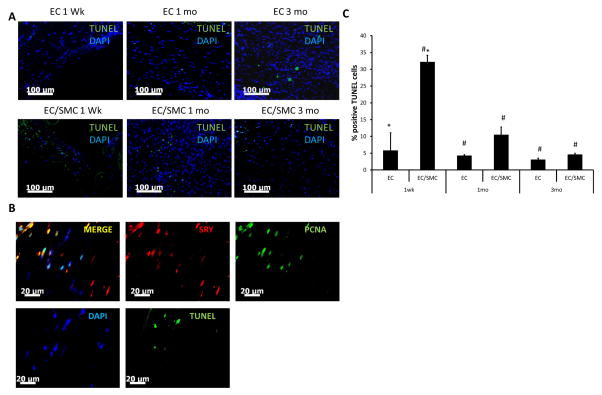
Figure 6. Evaluation of apoptotic cells within the vascular grafts.
(A) TUNEL assay was performed on paraffin sections of explanted TEVs. TUNEL positive nuclei appear green; nuclei were counterstained with DAPI (blue). Bar=100 μm. (B) Top panel: immunostaining of 1 wk explanted TEVs for SRY (red) and PCNA (green) (TOP panels). Bottom panel: TUNEL+ nuclei (green) in a consecutive tissue section; nuclei were counterstained with DAPI (blue). Bar= 20 μm. (C) Quantification of TUNEL+ nuclei as percentage of total (DAPI+) nuclei (#: p<0.02, *: p<0.05, n=3).

