
Fig. 4.
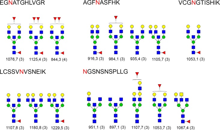
Proposed structures for Cubilin belonging to five different N-glycosylation sites. Blue squares are HexNAc, green circles are mannoses, and yellow circles are galactose, whereas the red triangle is fucose. The peptide sequence is shown at the top of each box. Five different peptide sequences are shown. N-glycosylation site is marked with a red letter. The m/z value of each precursor is given at the bottom of the glycan structure and the charge is given in parenthesis. When the antennalfucose position is ambiguous such as attached to either HexNAc or Hexose, then the fucose is drawn outside of the parenthesis. It is to be noted that LacNAc units cannot be assigned to a given branch from the spectrum; and therefore, the number of antennae remains ambiguous. Here, the structures from GlycomeDB that matched to various spectrums are shown. When the glycan composition is determined, if there are several identical compositions in GlycomeDB with different structures the potential structures are theoretically fragmented and compared with the empirical spectrum and scored. The different structures yield different fragments and the highest scoring structure is reported.