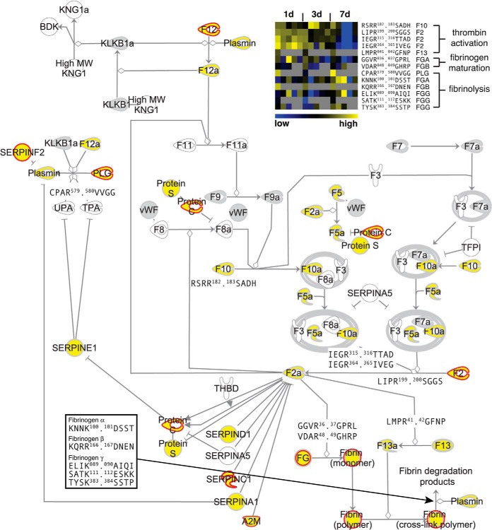
Fig. 4.
Proteolytic map of blood coagulation. Ingenuity pathway analysis was used to map proteins and N termini onto the canonical blood coagulation cascade. Proteins identified only by tryptic peptides are in gray, proteins with N-terminal annotation in yellow. Known processing events revealed by corresponding protein neo-N termini are shown by their P4-P4′ cleavage site motifs. Positions are related to unprocessed precursor sequences. A heatmap of relative abundances of N-terminal peptides derived from indicated cleavages in wound fluids at different time points after wounding is shown in the top right corner. Gray squares indicate missing values for replicate experiments. Neo-N termini generated during thrombin activation and fibrinogen maturation are higher abundant in fluids collected early and neo-N-terminal peptides from fibrin degradation (fibrinolysis) in samples obtained later after injury. Proteins predicted to be cleaved by MMPs (see Fig. 6A) are marked by red borders.

