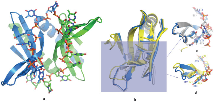
Figure 2. The overall structure of the PC4 mutant-ssDNA complex and the comparison of the structure of PC4 wild type or mutant DNA complexes with MoSub1 DNA-bound structure.
Overall structure of the PC4 mutant-ssDNA complex (a). Chains A-B, shown in cartoon representation, are coloured in blue and green respectively. Superposition of the monomer structure of PC4 wild type (grey), PC4 mutant (blue) and MoSub1 (yellow) DNA complex (b). The region around residue 74 or 89 is highlighted to show different DNA conformations in the PC4 mutant and PC4 wild type (c), PC4 mutant and MoSub1 (d) DNA complexes.

