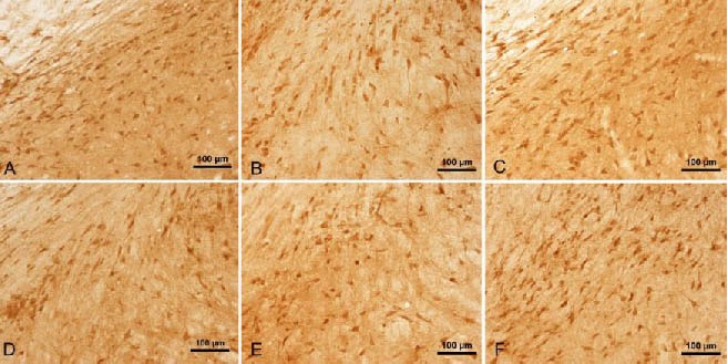
Figure 3.

Tumor necrosis factor-α staining in the midbrains of rats (immunohistochemical staining, light microscope, scale bars: 100 μm).
Many dark brown-colored cells can be observed in the control rats (A).
The number of positive cells was reduced in the model rats at 4 days (B) and 14 days (C). This was also the case in the gastrodin (D) and Madopar (E) groups at 14 days.
The number of positive cells in the Madopar group at 28 days (F) was greater than that at 14 days (E).
