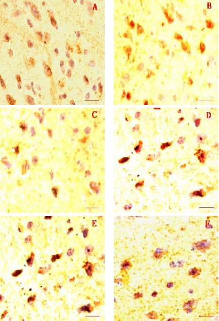
Figure 3.

Immunohistochemical staining of the substantia nigra by tyrosine hydroxylase. Scale bars: 2 µm.
(A) Normal control group; (B) Herba Epimedii group; (C) Fructus Ligustri Lucidi group; (D) Rhizoma Polygonati group; (E) selegiline group; (F) model group.
The number of tyrosine hydroxylase-positive cells in the Herba Epimedii group was similar with that in the normal control group, but that was higher than that in the Fructus Ligustri Lucidi, Rhizoma Polygonati, selegiline, and model groups.
