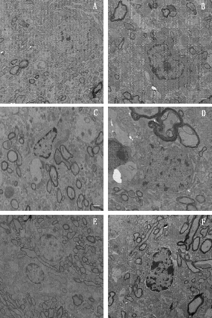
Figure 4.

Transmission electron microscopy images of substantia nigra neurons from each group. Scale bars: 2 µm.
(A) Normal control group; (B) Herba Epimedii group; (C) Fructus Ligustri Lucidi group; (D) Rhizoma Polygonati group; (E) selegiline group; (F) model group.
In the normal control group, an intact cell membrane and organelles were observed. In the Herba Epimedii, Fructus Ligustri Lucidi, Rhizoma Polygonati, selegiline, and model groups, pyknosis of different degrees and chromatin margination were observed.
